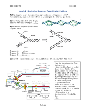
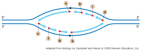
41 the diagram below shows a bacterial replication fork and its principal proteins.
Mastering Biology ch. 13 Flashcards | Quizlet In DNA replication in bacteria, the enzyme DNA polymerase III (abbreviated DNA pol III) adds nucleotides to a template strand of DNA. But DNA pol III cannot start a new strand from scratch. Instead, a primer must pair with the template strand, and DNA pol III then adds nucleotides to the primer, complementary to the template strand. Molecular mechanism of DNA replication - Khan Academy Illustration shows the replication fork. Helicase unwinds the helix, and single-strand binding proteins prevent the helix from re-forming. Topoisomerase prevents the DNA from getting too tightly coiled ahead of the replication fork. DNA primase forms an RNA primer, and DNA polymerase extends the DNA strand from the RNA primer.
Mastering Biology Chp. 13 HW Flashcards | Quizlet In DNA replication in bacteria, the enzyme DNA polymerase III (abbreviated DNA pol III) adds nucleotides to a template strand of DNA. But DNA pol III cannot start a new strand from scratch. Instead, a primer must pair with the template strand, and DNA pol III then adds nucleotides to the primer, complementary to the template strand.

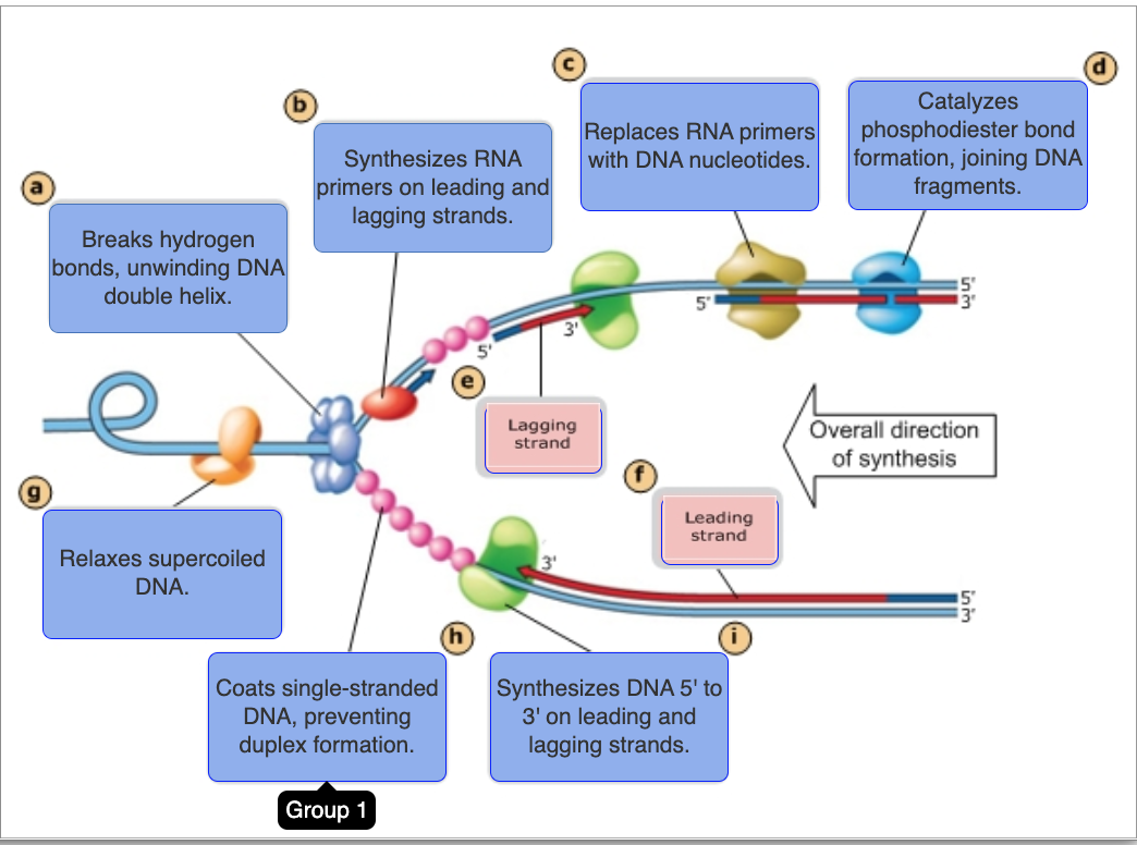
The diagram below shows a bacterial replication fork and its principal proteins.
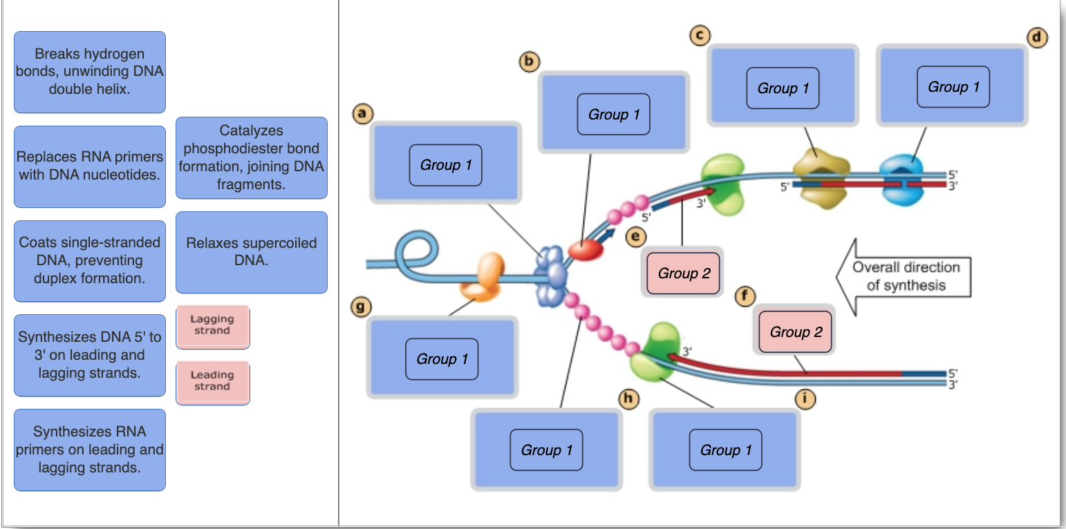
biology chapter 16 Flashcards | Quizlet Each of the four images below shows a strand of template DNA (dark blue) with an RNA primer (red) to which DNA pol III will add nucleotides. tcggccgt The diagram below shows a bacterial replication fork and its principal proteins. Drag the labels to their appropriate locations in the diagram to describe the name or function of each structure. Chapter 11 Flashcards | Quizlet The diagram below shows a bacterial replication fork and its principal proteins. Drag the labels to their appropriate locations in the diagram to describe the name or function of each structure. Use pink labels for the pink targets and blue labels for the blue targets. A) Breaks hydrogen bonds, unwinding DNA double helix. Solved The diagram below shows a bacterial replication fork ... Question: The diagram below shows a bacterial replication fork and its principal proteins. Drag the labels to their appropriate locations in the diagram to describe the name or function of each structure. Use pink labels for the pink targets and blue labels for the blue targets.
The diagram below shows a bacterial replication fork and its principal proteins.. Solved The diagram below shows a bacterial replication fork ... Question: The diagram below shows a bacterial replication fork and its principal proteins. Drag the labels to their appropriate locations in the diagram to describe the name or function of each structure. Use pink labels for the pink targets and blue labels for the blue targets. Chapter 11 Flashcards | Quizlet The diagram below shows a bacterial replication fork and its principal proteins. Drag the labels to their appropriate locations in the diagram to describe the name or function of each structure. Use pink labels for the pink targets and blue labels for the blue targets. A) Breaks hydrogen bonds, unwinding DNA double helix. biology chapter 16 Flashcards | Quizlet Each of the four images below shows a strand of template DNA (dark blue) with an RNA primer (red) to which DNA pol III will add nucleotides. tcggccgt The diagram below shows a bacterial replication fork and its principal proteins. Drag the labels to their appropriate locations in the diagram to describe the name or function of each structure.
Division of Labor between PCNA Loaders in DNA Replication and ...
Questions PS2 inital - Module 2 - Replication, Repair and ...

Solved Complete diagram of a replication fork in bacterial ...
Chapter 11: DNA Replication (Homework) Flashcards | Chegg.com

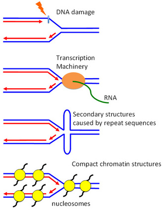
Linking RNA Polymerase Backtracking to Genome Instability in ...
Chapter 9: DNA Replication – Chemistry

Continuous Cell-Free Replication and Evolution of Artificial ...

DNA unwinding model. (A) Diagram showing notation used for ...

Chapter 13: molecular basis of inheritance Flashcards | Quizlet

Mastering Biology Chapter 16 – RHS Homework
Mastering Biology Chapter 16 – RHS Homework

Chapter 9: DNA Replication – Chemistry
Solved] Label the figure to assess your knowledge of DNA ...
Mastering Biology Chapter 16 – RHS Homework

Genetics test 3 Flashcards | Quizlet
RPA directs SMARCAL1 specifically to a damaged replication ...

Mastering Biology Chp. 13 HW Flashcards | Quizlet
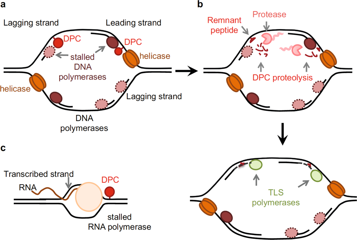
DNA–protein crosslink proteases in genome stability ...
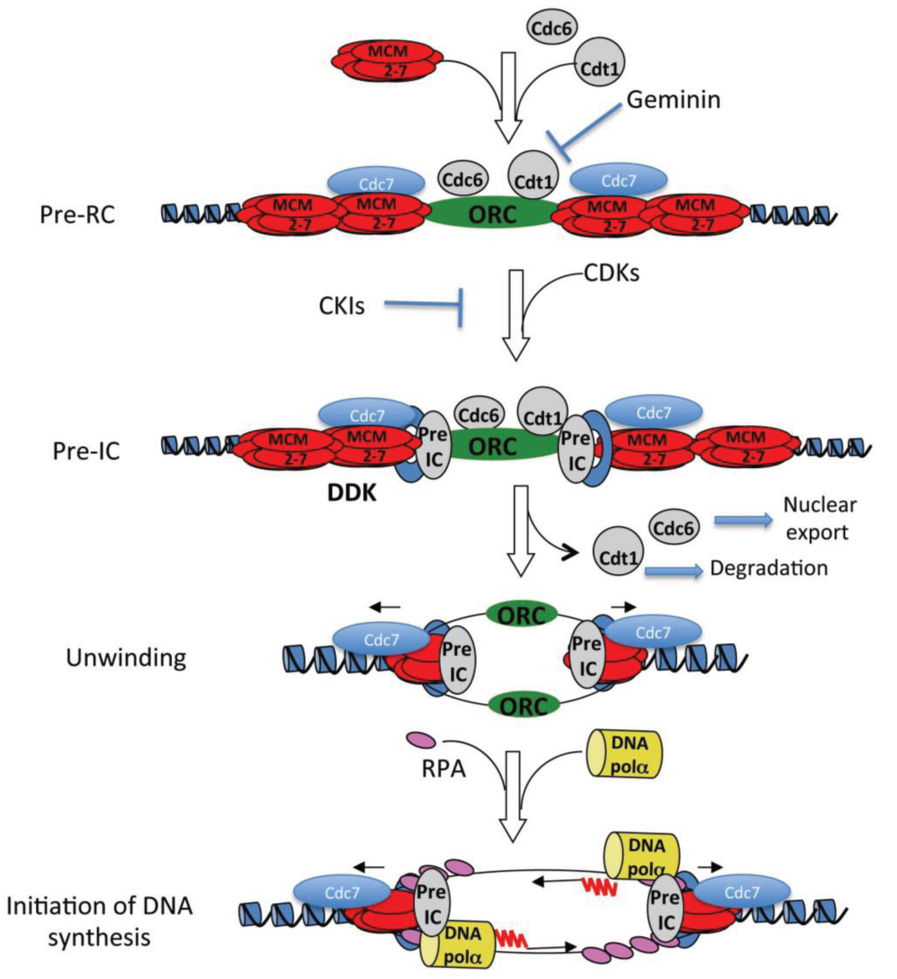
DNA Replication, Checkpoint, DNA Synthesis | Learn Science at ...

Biomolecules | Free Full-Text | DNA Methylation in Regulatory ...

New insights into replisome fluidity during chromosome ...

New Antimicrobials Targeting Bacterial RNA Polymerase ...

Mastering Biology Exam 3 Flashcards | Quizlet
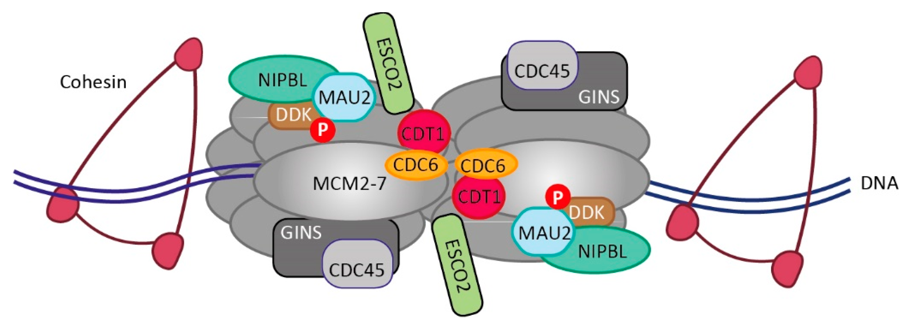
Cells | Free Full-Text | The Interplay of Cohesin and the ...

DNA primase acts as a molecular brake in DNA replication | Nature

Lecture 15 Post-Class Questions Adaptive Follow-Up Flashcards ...

Okazaki fragment maturation: DNA flap dynamics for cell ...

Chapter 12: DNA Damage and Repair – Chemistry
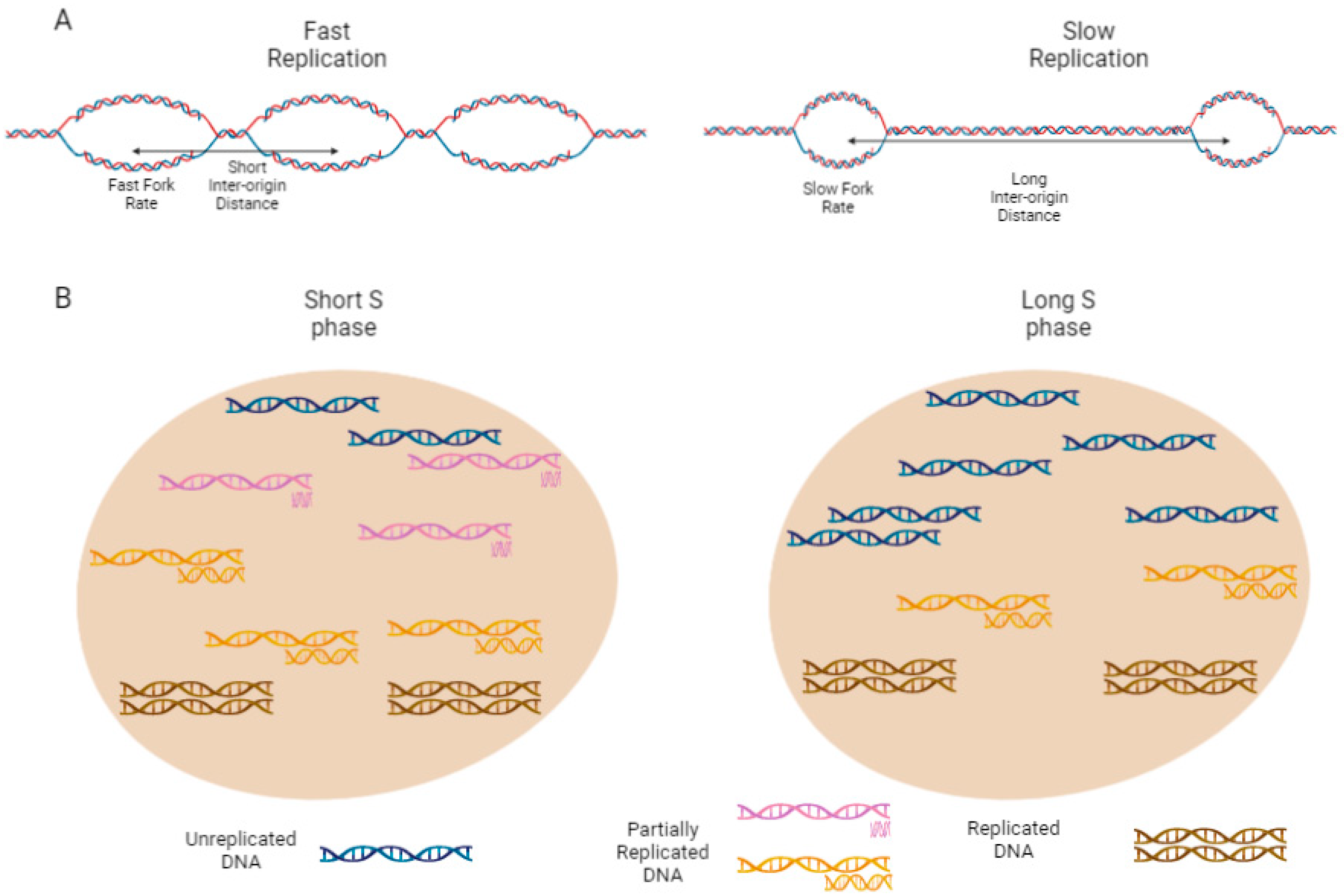
Biology | Free Full-Text | S Phase Duration Is Determined by ...

DNA Replication - Leading Strand vs Lagging Strand & Okazaki Fragments
Impeded DNA replication forks can be rescued by different ...

intro to cell test five Flashcards | Quizlet
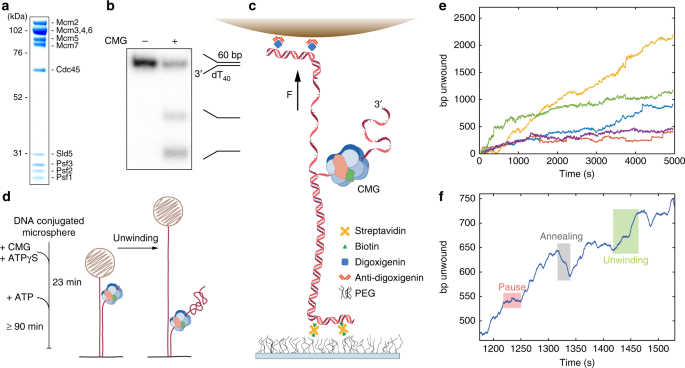
The mechanism of DNA unwinding by the eukaryotic replicative ...

Strand-Specific Analysis Shows Protein Binding at Replication ...
Chapter 11: DNA Replication (Homework) Flashcards | Chegg.com
What's the difference between gene replication and DNA ...
Genes | Free Full-Text | Molecular Mechanisms of DNA ...

Mastering Biology ch. 13 Flashcards | Quizlet
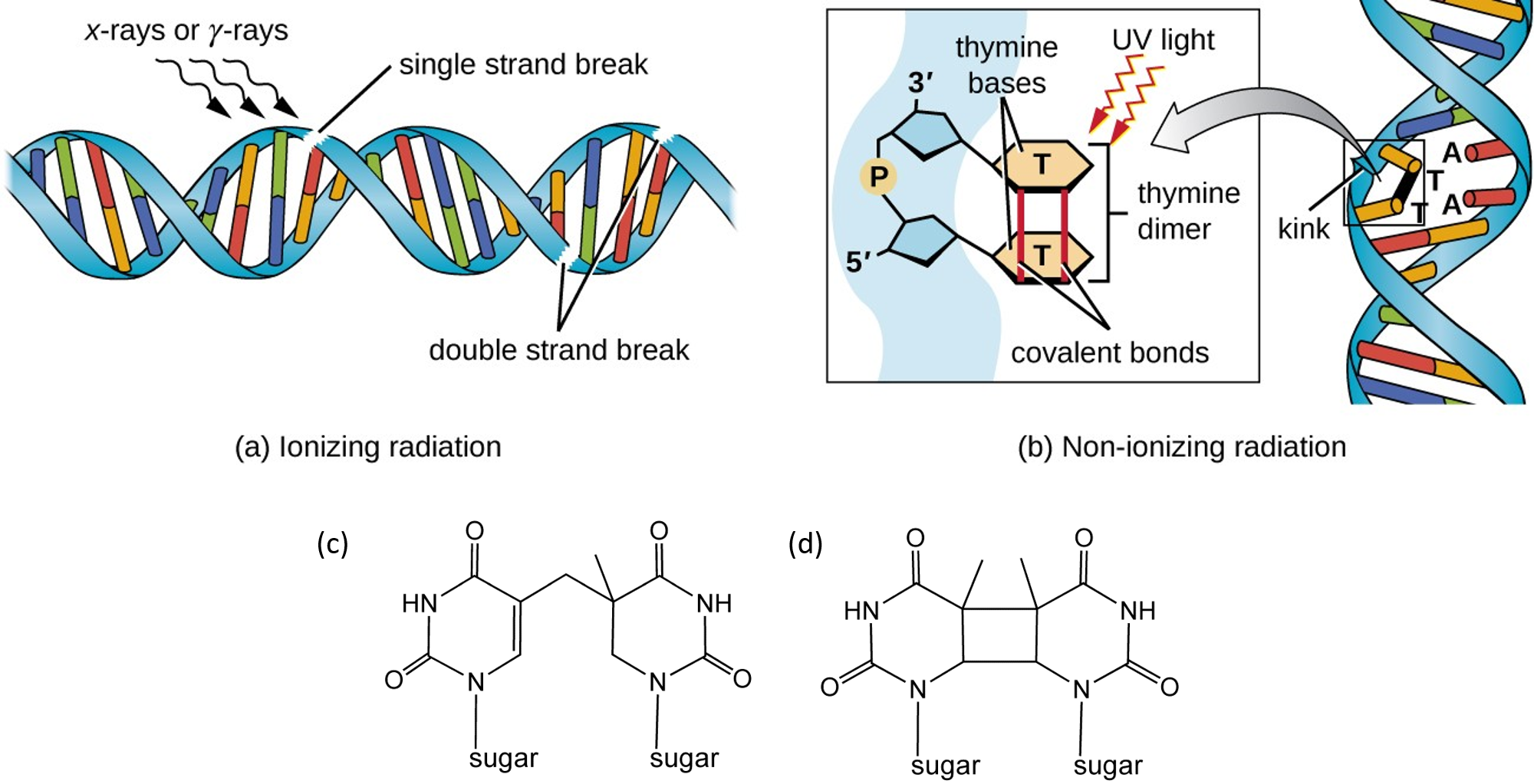
Chapter 12: DNA Damage and Repair – Chemistry

Genetics Exam 1 Test Questions Flashcards | Quizlet
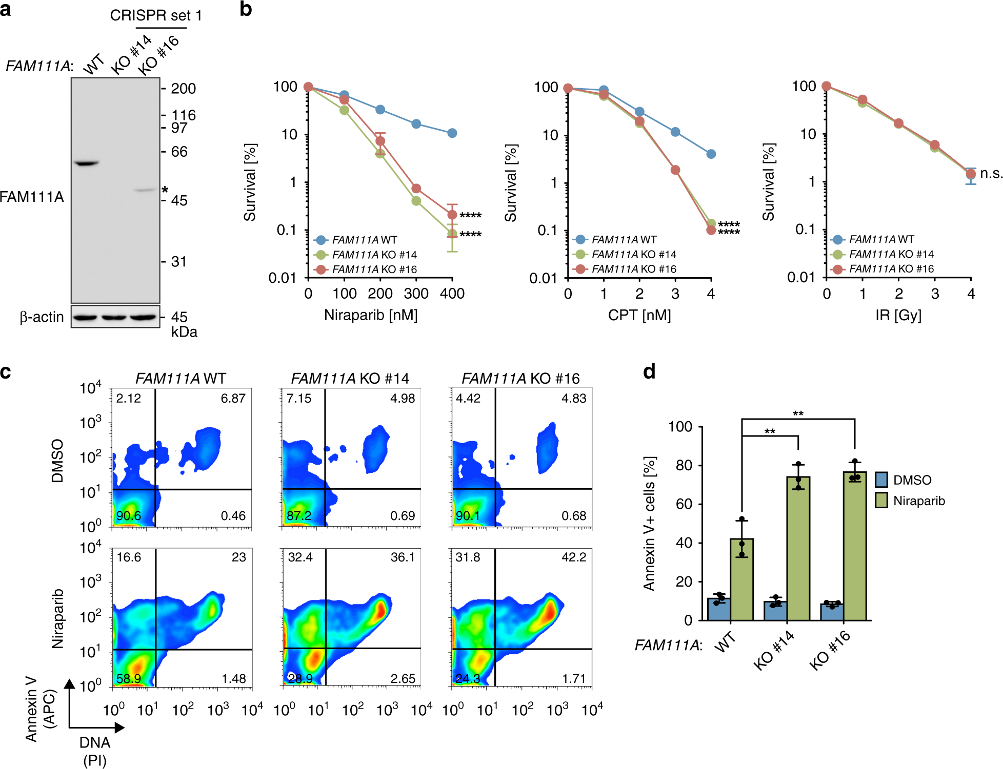
FAM111A protects replication forks from protein obstacles via ...
Post a Comment for "41 the diagram below shows a bacterial replication fork and its principal proteins."