44 labeled diagram of compound microscope
Compound Microscope: Definition, Diagram, Parts, Uses, Working ... - BYJUS The parts of a compound microscope can be classified into two: Non-optical parts Optical parts Non-optical parts Base The base is also known as the foot which is either U or horseshoe-shaped. It is a metallic structure that supports the entire microscope. Pillar The connection between the base and the arm are possible through the pillar. Arm Labeled Cell Leaf Microscope Under Search: Leaf Cell Under Microscope Labeled. She saw a cell part that could use energy from This is because most of You can find these cancer-causing, cannibal-style injections listed on the CDC Estimate cell size (if you have previously calibrated your microscope) Light & electron microscopes - preparation of samples for investigation e Light & electron microscopes - preparation of samples for ...
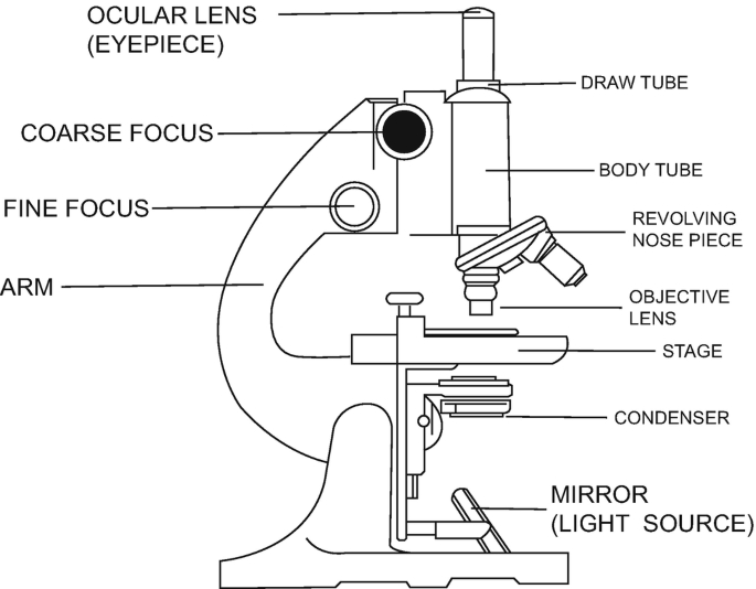
Labelled Diagram of Compound Microscope - Biology Discussion The below mentioned article provides a labelled diagram of compound microscope. Part # 1. The Stand: The stand is made up of a heavy foot which carries a curved inclinable limb or arm bearing the body tube. The foot is generally horse shoe-shaped structure (Fig. 2) which rests on table top or any other surface on which the microscope in kept.

Labeled diagram of compound microscope
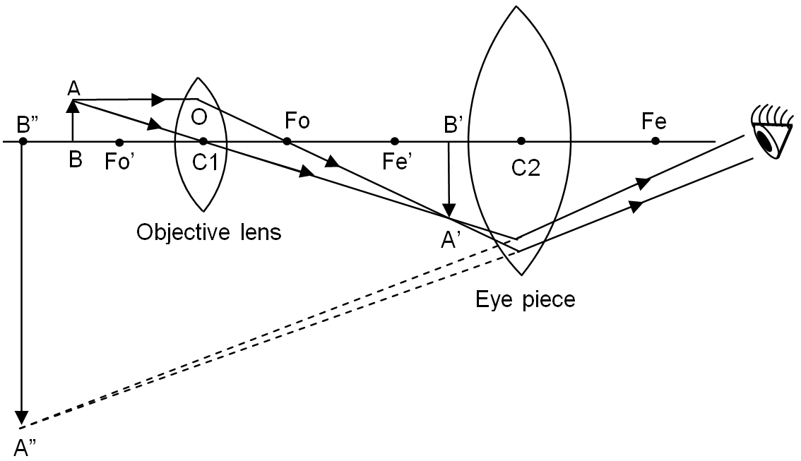
(b) Why both objective and eyepiece of a compound microscope must have ... Question (a) Draw the labelled ray diagram for the formation of image by a compound microscope. Derive an expression for its total magnification (or magnifying power), when the final image is formed at the near point. (b) Why both objective and eyepiece of a compound microscope must have short focal lengths? Diagram of a Compound Microscope - Biology Discussion The size of objects viewed under the compound microscope can be accurately determined using a micrometer. The latter consists of two scales, the eyepiece scale, (also called 'graticule' or 'ocular') and the stage micrometer scale. The eyepiece scale is calibrated with the help of stage micrometer and the former is then used for measurements. Binocular Microscope Anatomy - Parts and Functions with a Labeled Diagram Now, I will discuss the details anatomy of the light compound microscope with the labeled diagram. Why it is called binocular: because it has two ocular lenses or an eyepiece on the head that attaches to the objective lens, this ocular lens magnifies the image produced by the objective lens. Binocular microscope parts and functions
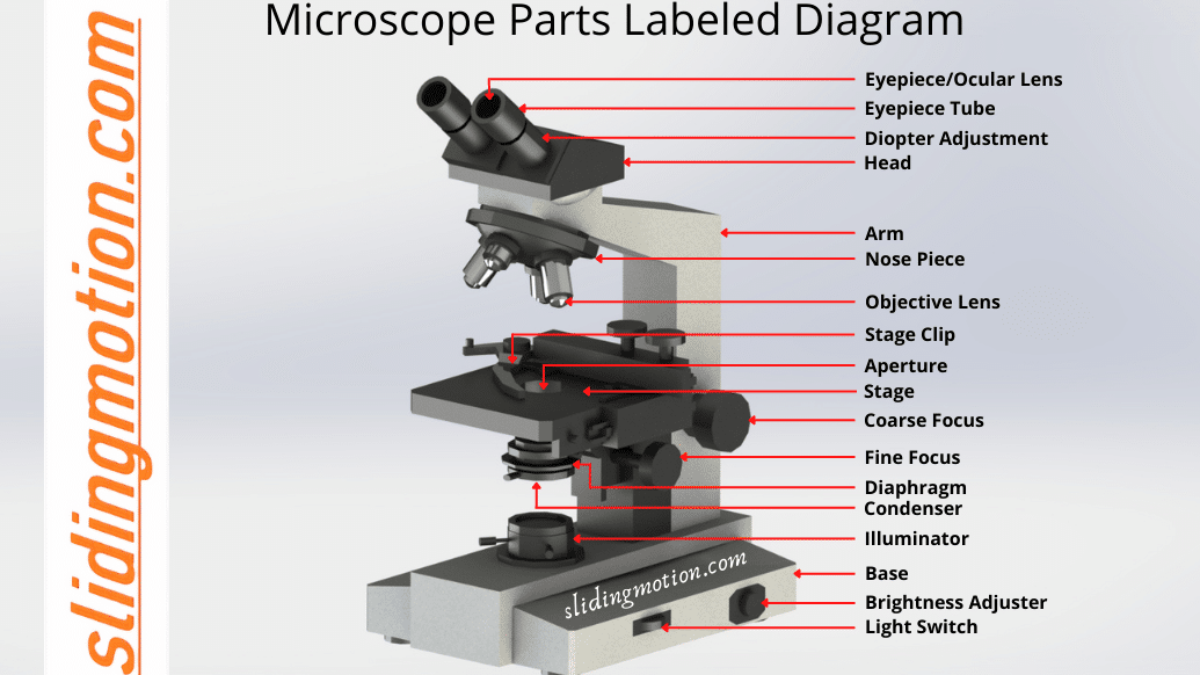
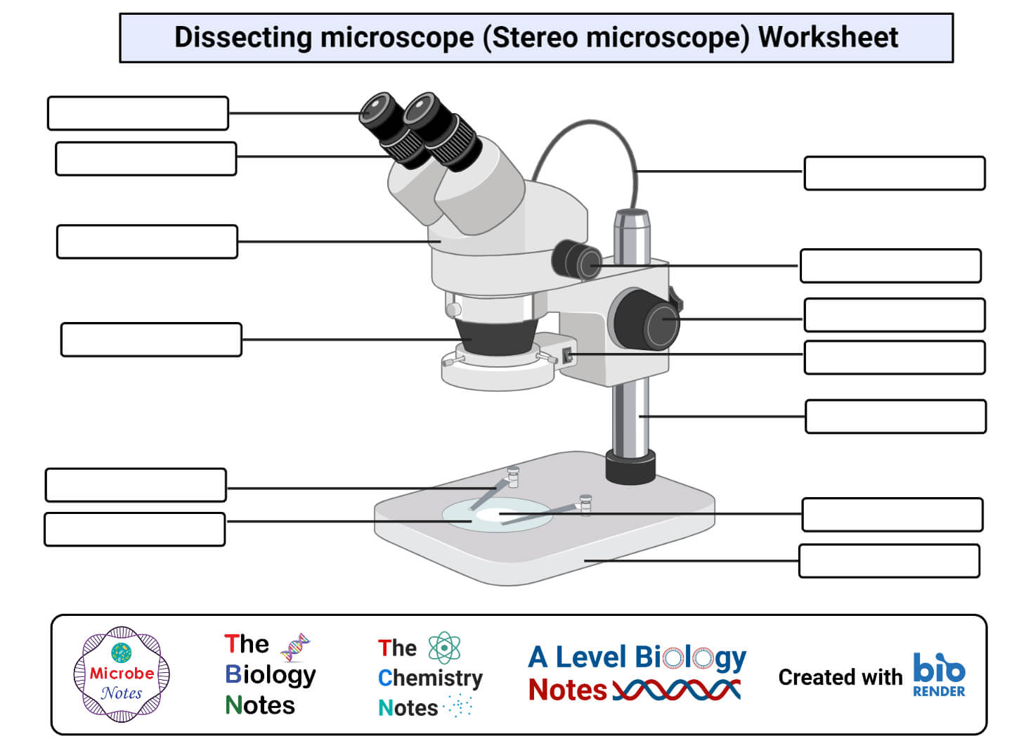
Labeled diagram of compound microscope. Parts of Stereo Microscope (Dissecting microscope) - labeled diagram ... If you would like to learn optical components of a compound microscope, please visit Compound Microscope Parts - Labeled Diagram and their Functions, and this article. How to use a stereo (dissecting) microscope. Follow these steps to put your stereo microscopes in work: 1. Set your microscope on a tabletop or other flat sturdy surface where ... Microscope Parts, Function, & Labeled Diagram - slidingmotion Microscope parts labeled diagram gives us all the information about its parts and their position in the microscope. Microscope Parts Labeled Diagram The principle of the Microscope gives you an exact reason to use it. It works on the 3 principles. Magnification Resolving Power Numerical Aperture. Parts of Microscope Head Base Arm Eyepiece Lens Compound Microscope Parts, Functions, and Labeled Diagram Compound Microscope Parts, Functions, and Labeled Diagram Parts of a Compound Microscope Each part of the compound microscope serves its own unique function, with each being important to the function of the scope as a whole. Draw a neat labelled diagram of a compound microscope and ... - Sarthaks Description : It consists of two convex lenses separated by a distance. The lens near the object is called objective and the lens near the eye is called eye piece. The objective lens has small focal length and eye piece has of larger focal length. The distance of the object can be adjusted by means of a rack and pinion arrangement.
And Parts Use Microscope Worksheet Quizlet Microscopy compound microscope parts of compound microscope parts parts of a compound microscope with labeled diagram and functions The electron microscope Clean your microscope slides with lens paper before examining Parts of a Microscope Use the completed résumé as a reference for filling out the application Serves as a handle to carry the ... 16 Parts of a Compound Microscope: Diagrams and Video Once you have an understanding of the parts of the microscope it will be much easier to navigate around and begin observing your specimen, which is the fun part! The 16 core parts of a compound microscope are: Head (Body) Arm Base Eyepiece Eyepiece tube Objective lenses Revolving Nosepiece (Turret) Rack stop Coarse adjustment knobs Label a Compound Microscope Diagram | Quizlet Only $2.99/month Label a Compound Microscope STUDY Learn Flashcards Write Spell Test PLAY Match Gravity Created by Hesi_Study Terms in this set (16) Label this Eyepiece (ocular lens) Label this Body tube Label this Arm Label this Mechanical Stage Control Knobs Label this Coarse Adjustment Knob Label this Fine Adjustment Knob Label this Base Parts of a Compound Microscope and Their Functions - NotesHippo Compound microscope mechanical parts (Microscope Diagram: 2) include base or foot, pillar, arm, inclination joint, stage, clips, diaphragm, body tube, nose piece, coarse adjustment knob and fine adjustment knob.. Base: It's the horseshoe-shaped base structure of microscope.All of the other components of the compound microscope are supported by it. ...
Compound Microscope - Diagram (Parts labelled), Principle and Uses See: Labeled Diagram showing differences between compound and simple microscope parts Structural Components The three structural components include 1. Head This is the upper part of the microscope that houses the optical parts 2. Arm This part connects the head with the base and provides stability to the microscope. Compound Microscope Parts, Function, & Diagram - Study.com Learn the compound light microscope's parts and functions by viewing a compound microscope diagram. Also, read about the uses of a compound microscope. Updated: 11/04/2021 Compound Microscope Labeled Diagram | Quizlet QUESTION. The total magnification of a specimen being viewed with a 10X ocular lens and a 40X objective lens is. 15 answers. QUESTION. a mosquito beats its wings up and down 600 times per second, which you hear as a very annoying 600 Hz sound. if the air outside is 20 C, how far would a sound wave travel between wing beats. 2 answers. Compound Microscope: Parts of Compound Microscope - BYJUS Diagram Parts of the Compound Microscope Parts of Compound Microscope The parts of the compound microscope can be categorized into: Mechanical parts Optical parts (A) Mechanical Parts of a Compound Microscope 1. Foot or base It is a U-shaped structure and supports the entire weight of the compound microscope. 2. Pillar It is a vertical projection.
Parts of Stereo Microscope (Dissecting microscope) – labeled ...
Microscope Types (with labeled diagrams) and Functions Compound microscope labeled diagram Compound microscope functions: It finds great application in areas of pathology, pedology, forensics etc Its greater order of magnification allows for deeper study of microbial organisms to Detect the cause of diseases Study the mineral composition in soils

Compound light microscope parts, Magnification formula
Compound Microscope Parts - Labeled Diagram and their Functions Labeled diagram of a compound microscope Major structural parts of a compound microscope There are three major structural parts of a compound microscope. The head includes the upper part of the microscope, which houses the most critical optical components, and the eyepiece tube of the microscope.

How to draw compound of Microscope easily - step by step
PDF Labelled Microscope Diagrams Of The Human Testes April 27th, 2018 - With Labeled Diagram and Functions How does a Compound Microscope Work Before exploring the parts of a compound microscope you should probably understand that the compound light microscope is more complicated than just a microscope with more

Compound Microscope Parts, Functions, and Labeled Diagram ...
(a) Draw a labelled ray diagram of compound microscope, when final ... (a) Draw a labelled ray diagram of compound microscope, when final image forms at the least distance of distinct vision. (b) Why is its objective of short focal length and of short aperture, compared to its eyepiece? Explain. (c) The focal length of the objective is 4 cm while that of eyepiece is 10 cm.

What is a Compound Microscope? | Microscope World Blog
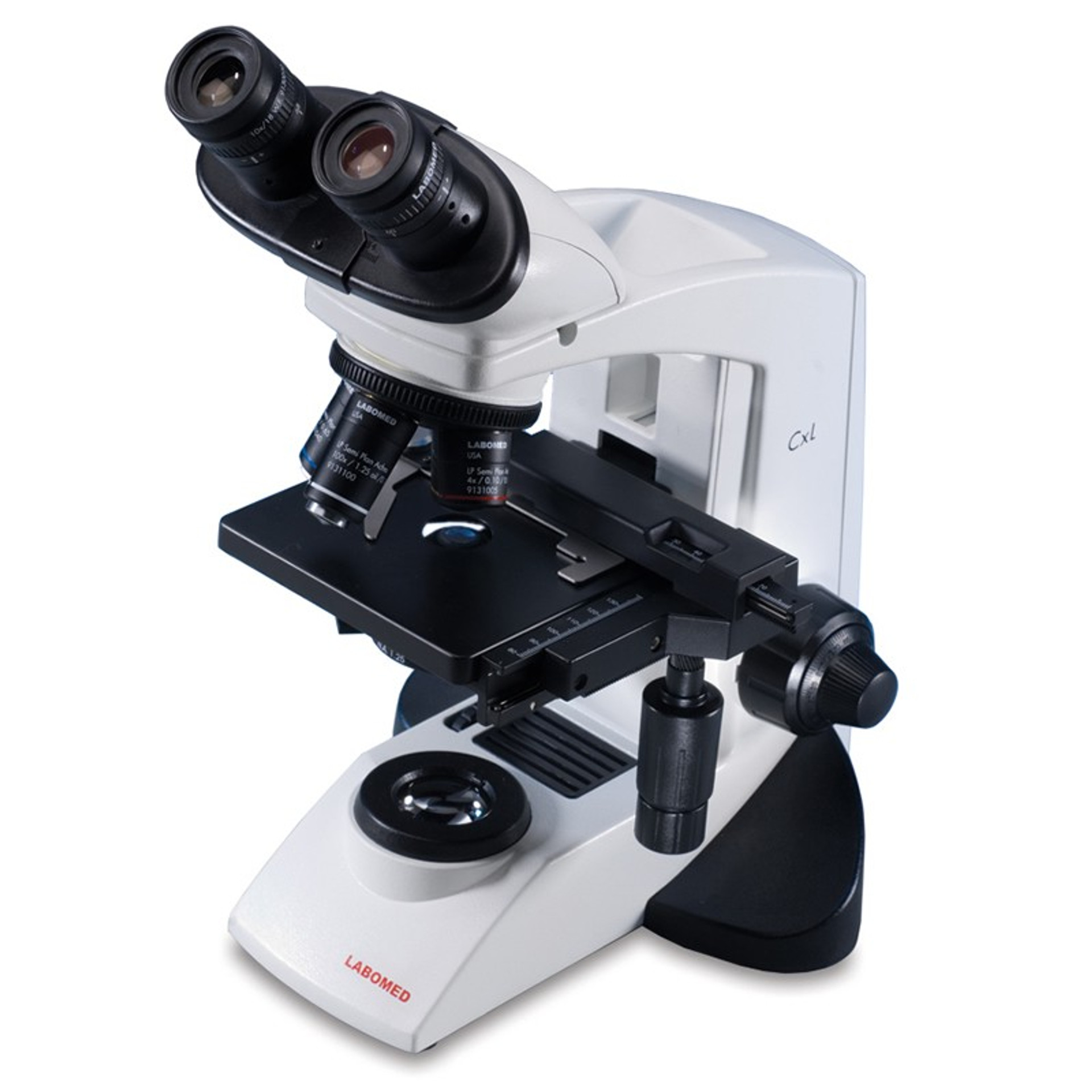
Compound Microscope - Types, Parts, Diagram, Functions and Uses It comes with a wide body and base. Its distinct parts include a condenser, illumination, focus lock, mechanical stage, and a revolving nosepiece which can hold up to five objectives. It usually has a binocular head, which makes long-term observation easy. Image 22: An example of a research compound microscope.
Parts of Microscope, Function, Names & Labeled Diagram ...
Parts of a microscope with functions and labeled diagram - Microbe Notes Q. List down the 18 parts of a Microscope. 1. Ocular Lens (Eye Piece) 2. Diopter Adjustment 3. Head 4. Nose Piece 5. Objective Lens 6. Arm (Carrying Handle) 7. Mechanical Stage 8. Stage Clip 9. Aperture 10. Diaphragm 11. Condenser 12. Coarse Adjustment 13. Fine Adjustment 14. Illuminator (Light Source) 15. Stage Controls 16. Base 17.

Compound Microscope Parts, Functions, and Labeled Diagram ...
biology diagram full hd 31 Diagram Of Amoeba With Label - Labels Database 2020 ardozseven.blogspot.com. amoeba labeled spirogyra biology diagram label paramecium protista functions malaria. ... Unlabelled Diagram Of Compound Microscope - Micropedia microspedia.blogspot.com. unlabelled 40x 100x 3000f dissecting. Science Backgrounds - WallpaperSafari ...

This is a common compound microscope. Label its parts from A ...
Microscope Parts and Functions Most specimens are mounted on slides, flat rectangles of thin glass. The specimen is placed on the glass and a cover slip is placed over the specimen. This allows the slide to be easily inserted or removed from the microscope. It also allows the specimen to be labeled, transported, and stored without damage.
Exercise 1: Using a Compound Microscope | SpringerLink
(a) Draw a labelled ray diagram of a compound microscope. (b) Derive an ... Best answer (a) Labelled diagram of compound microscope. The objective lens form image A' B' near the first focal point ofeyepiece. (b) Angular magnification of objective lens m0 = linear magnification h'/h where L is the distance between second focal point of the objective and first focal point of eyepiece.

Compound Microscope: Parts of Compound Microscope
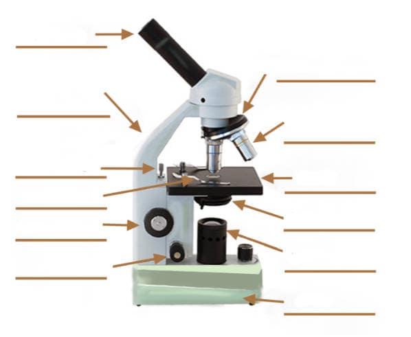
A Study of the Microscope and its Functions With a Labeled Diagram ... These labeled microscope diagrams and the functions of its various parts, attempt to simplify the microscope for you. However, as the saying goes, 'practice makes perfect', here is a blank compound microscope diagram and blank electron microscope diagram to label. Download the diagrams and practice labeling the different parts of these ...
Parts of the Microscope with Labeling (also Free Printouts ...
Compound Microscope- Definition, Labeled Diagram, Principle, Parts, Uses The naked eye can now view the specimen at magnification 400 times greater and so microscopic details are revealed. Alternatively, the magnification of the compound microscope is given by: m = D/ fo * L/fe where, D = Least distance of distinct vision (25 cm) L = Length of the microscope tube fo = Focal length of the objective lens

Microscope With Labels Clip Art at Clker.com - vector clip ...
Binocular Microscope Anatomy - Parts and Functions with a Labeled Diagram Now, I will discuss the details anatomy of the light compound microscope with the labeled diagram. Why it is called binocular: because it has two ocular lenses or an eyepiece on the head that attaches to the objective lens, this ocular lens magnifies the image produced by the objective lens. Binocular microscope parts and functions

Compound Microscope Parts – Labeled Diagram and their ...
Diagram of a Compound Microscope - Biology Discussion The size of objects viewed under the compound microscope can be accurately determined using a micrometer. The latter consists of two scales, the eyepiece scale, (also called 'graticule' or 'ocular') and the stage micrometer scale. The eyepiece scale is calibrated with the help of stage micrometer and the former is then used for measurements.

compound microscope Diagram | Quizlet
(b) Why both objective and eyepiece of a compound microscope must have ... Question (a) Draw the labelled ray diagram for the formation of image by a compound microscope. Derive an expression for its total magnification (or magnifying power), when the final image is formed at the near point. (b) Why both objective and eyepiece of a compound microscope must have short focal lengths?

File:Labelledmicroscope.gif - Wikimedia Commons
Compound Microscope Parts, Functions, and Labeled Diagram ...

Label the Microscope Diagram | Download Scientific Diagram

Parts of a Microscope with Their Functions • Microbe Online
The Compound Light Microscope Label the following parts on ...
Compound Microscope Drawing - ClipArt Best
Parts of a microscope with functions and labeled diagram

Draw a labelled diagram of a compound microscope.

draw the labelled ray diagram for the formation of image by a ...
a) Draw a labelled ray diagram of a compound microscope. (b ...
Draw a neat labelled diagram of a compound microscope and ...

Compound Microscope Parts – Labeled Diagram and their ...

Label Microscope Diagram - EnchantedLearning.com

Parts of Microscope, Function, Names & Labeled Diagram ...
Draw a labelled ray diagram of a compound microscope and ...

2.1 " Compound Microscope" | Download Scientific Diagram
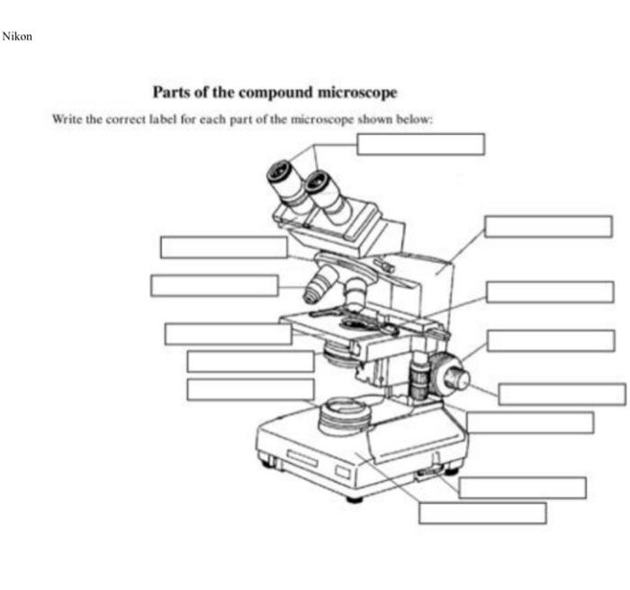
Solved Nikon Parts of the compound microscope Write the ...

Compound Light Microscope Labeling Diagram | Quizlet

microscope diagram with name - EDUSIP

Compound Microscope Parts – Labeled Diagram and their ...

Compound Microscope Parts, Functions, and Labeled Diagram ...

Microscope labeled diagram

Draw a labelled diagram of a compound microscope.

This is a common compound microscope. Label its parts from A ...

Microscope Labeling Diagram | Quizlet
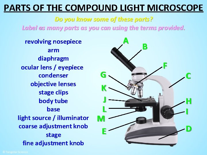
MICROSCOPE PARTS PARTS OF THE COMPOUND LIGHT MICROSCOPE

Compound Microscope Parts

Lesson Explainer: Microscopy | Nagwa

Labeling the Parts of the Microscope | Microscope World Resources

Compound and Stereo Microscopes | Download Scientific Diagram
Post a Comment for "44 labeled diagram of compound microscope"