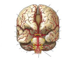
44 circle of willis without labels
Circle of Willis quizzes and unlabeled diagrams | Kenhub Get quizzing with our circle of willis quiz (arteries of the brain) below! On this circle of Willis quiz, you can choose from basic identification, advanced identification, clinical question banks and even intelligent mix - a personalised mix of all three. Central nervous system (CNS): Introduction to the brain Explore study unit. CT head sagittal - labeling questions - Radiopaedia.org Normal CT head (without labels) CT Loading Image 1 CT Sagittal non-contrast The same normal brain CT without labels for reference. Case Discussion The labeled structures are (excluding the correct side): mastoid air cells temporalis muscle zygomatic arch external auditory (acoustic) canal temporomandibular joint tentorium cerebelli sylvian fissure
(PDF) Anatomical labeling of the circle of willis using maximum a ... This structure is highly variable: only 42% of the population has a complete circle [2] and in the other cases, one or more arteries are missing. In the past years, several research papers addressed the problem of segmenting the CoW and the cerebral vasculature in general, without labeling them.

Circle of willis without labels
Cerebral Arterial Circle (Circle of Willis) - Neurosurgical Atlas Cerebral arterial circle (Circle of Willis). The brain has been removed to demonstrate an intact cerebral arterial circle that is formed by anastomotic connections between the basilar and internal carotid arteries. The basilar artery, formed by the union of the right and left vertebral arteries, terminates with the posterior cerebral arteries. Brain - Circle of Willis | Circle of willis, Willis, Circle This page presents a comprehensive series of labeled axial, sagittal and coronal images from a normal human brain magnetic resonance imaging exam. This MRI brain cross-sectional anatomy tool serves as a reference atlas to guide radiologists and researchers in the accurate identification of the brain structures. Melissa Pettyjohn vascular/other US 13.3 Circulation and the Central Nervous System - OpenStax The circle of Willis is a specialized arrangement of arteries that ensure constant perfusion of the cerebrum even in the event of a blockage of one of the arteries in the circle. The animation shows the normal direction of flow through the circle of Willis to the middle cerebral artery.
Circle of willis without labels. Brain Aneurysm Symptoms, Causes, Definition & Treatment Four major blood vessels supply blood to the brain. They join together at the Circle of Willis at the base of the brain. Smaller arteries leave the circle and branch out to supply brain cells with oxygen and nutrients. Artery junction points may become weak, causing a ballooning of the blood vessel wall to potentially form a small sac or aneurysm. Solved 18 possible...without plagiarizing 1) Some small | Chegg.com Ans 1) Circle of willis or cerebral arterial circle is a anostomotic ring of arteries formed between 2 vertebral and 2 internal carotid arteries which supply brain at the base of brain .It is formed by ; Posterior cerebral artery Posterior communicat… View the full answer A&P2 Lab - Circle of Willis Lab Flashcards | Quizlet circle of Willis The anastomotic polygon at the base of the brain, consisting of parts of the internal carotid, ACA, and PCA, interconnected by the anterior and posterior communicating arteries. anterior inferior cerebellar artery (AICA) A long, circumferential branch of the basilar artery arising just above the union of the 2 vertebrals. Variations in the Circle of Willis in a large population sample using ... These arteries anastomose to form the Circle of Willis (CoW) at the base of the brain (S1 Fig in S1 File ). The circular arrangement of the arteries enables the redistribution of blood flow when arteries in or upstream of the CoW experience reduced flow. This collateral ability of the CoW provides redundancy in the blood supply to the brain.
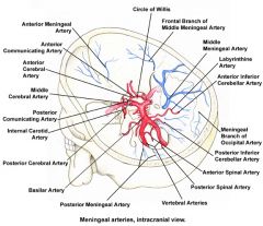
Circle of Willis - Wikipedia The circle of Willis (also called Willis' circle, loop of Willis, cerebral arterial circle, and Willis polygon) is a circulatory anastomosis that supplies blood to the brain and surrounding structures in reptiles, birds and mammals, including humans. [1] It is named after Thomas Willis (1621-1675), an English physician. [2] Contents 1 Structure File:Circle of Willis pt.svg - Wikipedia Better draw (more realistic). Bigger font size. Labels in colors. Added labels: Recurrent artery of Heubner and posteromedial central arteries, Circle of Willis, A1, A2, P1, P2. Correct arising for labyrinthine arteries. Use of layers. 20:33, 24 January 2007: 471 × 750 (39 KB) PbBR8498 CT angiography of the cerebral arteries (protocol) - Radiopaedia CT angiography of the cerebral arteries (also known as a CTA carotids or an arch to vertex angiogram) is a noninvasive technique allows visualization of the internal and external carotid arteries and vertebral arteries and can include just the intracranial compartment or also extend down to the arch of the aorta. Circle of Willis: Anatomy and function - Kenhub The circle of Willis is located on the inferior surface of the brain within the interpeduncular cistern of the subarachnoid space.It encircles various structures within the interpeduncular fossa (depression at the base of the brain) including the optic chiasm and infundibulum of the pituitary gland.. Although significant anatomic variations exist, the circle of Willis is typically composed of ...
brain MRA protocols and planning | indications for magnetic resonance ... 3D time-of-flight (TOF) Plan the axial 3D block on the sagittal plane; angle the position block parallel to the genu and splenium of the corpus callosum.Slices must be sufficient to cover the whole Circle of Willis from the corpus callosum up to the line of the foramen magnum. Check the positioning block in the other two planes. 1. The Cranial Nerves and the Circle Of Willis The Cranial Nerves and the Circle Of Willis Revised August 2, 2007 The objectives of this chapter are to identify: The structural divisions of the central nervous system (CNS). The cranial nerve roots associated with each brain stem division. The connective tissue wrappings, or the meninges, of the brain and spinal cord. File:Circle of Willis fr.svg - Wikimedia Commons Better draw (more realistic). Bigger font size. Labels in colors. Added labels: Recurrent artery of Heubner and posteromedial central arteries, Circle of Willis, A1, A2, P1, P2. Correct arising for labyrinthine arteries. Use of layers. 20:50, 30 May 2021: 480 × 756 (62 KB) Jmarchn (talk | contribs) Better draw (more realistic). Bigger font size. Neuroanatomy, Circle of Willis - StatPearls - NCBI Bookshelf The circle of Willis (CoW) is an anatomical structure that provides an anastomotic connection between the anterior and posterior circulations, providing collateral flow to affected brain regions in the event of arterial incompetency. The blood supply to the brain divides into an anterior and posterior circulation.
Circle Of Willis - Brain Blood Supply - YouTube
Anatomical Labeling of the Circle of Willis Using Maximum A Posteriori ... Thus, automating it becomes crucial for stream- T HE Circle of Willis (CoW) is a ring of cerebral arteries, located at the base of the brain, that connects the left and right anterior circulation with each other and with the posterior lining the geometric characterization of a large number of cases.
34 Label The Branches Of The Abdominal Aorta - Best Labels Ideas 2020
Variations in the Circle of Willis in a large population sample using ... The main arteries that supply blood to the brain originate from the Circle of Willis (CoW). The CoW exhibits considerable anatomical variations which may have clinical importance, but the variability is insufficiently characterised in the general population. We assessed the anatomical variability of CoW variants in a community-dwelling sample (N = 1,864, 874 men, mean age = 65.4, range 40-87 ...

RiT radiology: Intracranial Aneurysms
PDF IMAGING & DIAGNOSTICS CTA and the Circle of Willis group without history of CVA and with a normal CT brain postcontrast. There were 71 patients included in the stroke group with a history of recent and remote stroke and/or findings of stroke on CT scan. As Table 1 shows, 25 patients in the nonstroke group and 13 in the stroke group had a complete COW (P = .85) (Figure 2).
DITT Step 2 Day 1 Neuro/Psych Flashcards - Cram.com
The Circle Of Willis High-Res Vector Graphic - Getty Images Without a license, no further use can be made, such as: focus group presentations external presentations final materials distributed inside your organization any materials distributed outside your organization any materials distributed to the public (such as advertising, marketing)

Pin by Briana Lashaye on Health | Diagnostic medical sonography ...
File:Circle of Willis es.svg - Wikimedia Commons Size of this PNG preview of this SVG file: 380 × 599 pixels. Other resolutions: 152 × 240 pixels | 305 × 480 pixels | 487 × 768 pixels | 650 × 1,024 pixels | 1,300 × 2,048 pixels | 480 × 756 pixels. Original file (SVG file, nominally 480 × 756 pixels, file size: 61 KB) File information. Structured data.
DITT Step 2 Day 1 Neuro/Psych Flashcards - Cram.com
Circle of Willis: Anatomy, function, and what to know The circle of Willis is a junction of several important arteries at the bottom part of the brain. It helps blood flow from both the front and back sections of the brain. The circle of Willis gets...
Post a Comment for "44 circle of willis without labels"