44 diagram of an eye with labels
Labeled Eye Diagram | Eye anatomy diagram, Eye anatomy, Diagram of the eye accessory structures of the eye, extrinsic eye muscles, anatomy of the eyeball and microscopic anatomy of the retina. The skeletal system consists of bones and their associated connective tissues, including cartilage, tendons, and ligaments. It consists of dynamic, living tissues that are capable of growth, detect pain stimuli, adapt to stress ... Structure and Functions of Human Eye with labelled Diagram Structure and Functions of Human Eye with labelled Diagram Biology Biology Article Structure Of Eye Structure of the Eye The eye is one of the sensory organs of the body. In this article, we shall explore the anatomy of the eye The structure of the eye is an important topic to understand as it one of the important sensory organs in the human body.
The Human Eye (Eyeball) Diagram, Parts and Pictures The wall of the eyeball is made up of three layers - fibrous (outer), vascular/muscular (middle) and sensorineural (inner) layers. Diagram of the different layers of the eyeball Outer Layer The outer fibrous layer maintains the shape of the eyeball and protects more fragile internal structure. This layer is made up of the sclera and cornea.
Diagram of an eye with labels
Anatomy of the Eye Diagrams for Coloring/Labeling, with Reference and ... This printable contains 13 clear and simple cross sectional diagrams of the human eye. They photocopy well and are great for use as a labeling and coloring exercise for your students. The core eye anatomy diagram, designed as the labeling exercise, has a fully colored and labeled reference chart to go with it. Labelled Diagram of Human Eye, Explanation and Function - VEDANTU The human eye is a part of the sensory nervous system. Labeled Diagram of Human Eye The eyes of all mammals consist of a non-image-forming photosensitive ganglion within the retina which receives light, adjusts the dimensions of the pupil, regulates the availability of melatonin hormones, and also entertains the body clock. Eye Diagram With Labels and detailed description - BYJUS A brief description of the eye along with a well-labelled diagram is given below for reference. Well-Labelled Diagram of Eye The anterior chamber of the eye is the space between the cornea and the iris and is filled with a lubricating fluid, aqueous humour. The vascular layer of the eye, known as the choroid contains the connective tissue.
Diagram of an eye with labels. Labelling the eye — Science Learning Hub In this interactive, you can label parts of the human eye. Use your mouse or finger to hover over a box to highlight the part to be named. Drag and drop the text labels onto the boxes next to the eye diagram If you want to redo an answer, click on the box and the answer will go back to the top so you can move it to another box. Human Eye Diagram, How The Eye Work -15 Amazing Facts of Eye The shark has even been used in human eye surgery! FACT 4 The length of our eyes are about 1 inch across and weigh about 0.25 ounce. FACT 5 Our eyeballs stay the same size forever but our nose and ears continue to grow. FACT 6 Eyes are the second most complex organ after the brain. Generate eye diagram - MATLAB eyediagram - MathWorks eyediagram(x,n) generates an eye diagram for signal x, plotting n samples in each trace. The labels on the horizontal axis of the diagram range between –1/2 and 1/2. The function assumes that the first value of the signal and every nth value thereafter, occur at integer times. File:Diagram of human eye without labels.svg - Wikipedia File:Diagram of human eye without labels.svg. Size of this PNG preview of this SVG file: 410 × 430 pixels. Other resolutions: 229 × 240 pixels | 458 × 480 pixels | 732 × 768 pixels | 976 × 1,024 pixels | 1,953 × 2,048 pixels.
Label Eye Printout - EnchantedLearning.com Label the Eye Diagram. Human Anatomy. Read the definitions, then label the eye anatomy diagram below. Cornea - the clear, dome-shaped tissue covering the front of the eye. Iris - the colored part of the eye - it controls the amount of light that enters the eye by changing the size of the pupil. Lens - a crystalline structure located just behind ... Labeled Eye Diagram | Science Trends What you want to interpret as a major part of the human eye is somewhat up to the individual, but in general there are seven parts of the human eye: the cornea, the pupil, the iris, the lens, the vitreous humor, the retina, and the sclera. Let's take a closer look at each of these components individually. The Cornea Eye Anatomy: Parts of the Eye and How We See Behind the anterior chamber is the eye's iris (the colored part of the eye) and the dark hole in the middle called the pupil. Muscles in the iris dilate (widen) or constrict (narrow) the pupil to control the amount of light reaching the back of the eye. Directly behind the pupil sits the lens. The lens focuses light toward the back of the eye. The Anatomy of Human Eye with Diagram | EdrawMax Online 1. The Anatomy of Human Eye. The most complex sensory organs of the human body are the eyes. Every part of the human body is responsible for a specific action, from the muscles and tissues to the nerves and the blood vessels. The human eye consists of many muscles and tissues that join to form an approximately spherical structure.
Www Drawing Of Eye With There Label Labeled picture of the human eye. The eye is linked together with the nervous system which allows the brain to take in information from the eyes and make the appropriate decisions on how to act upon this information. Label Parts of the Human Eye. The eye is the organ responsible for our sense of sight. Drawing an Eye with only one Pencil. Module 1: Labeled Diagram of the Eye | Diagram of the eye, Eye anatomy ... Visual Field Test info, a visual field acuity test is a painless test that maps central vision as well as peripheral (side) vision. It is used to detect areas of vision loss (blind spots) caused by a brain tumor, stroke, glaucoma, diabetes, hypertension, or head trauma. pcasupportusa. P. Posterior Cortical Atrophy support USA. Car & Truck Body Part Diagrams | Auto Body Panel Names Mar 23, 2018 · Header panels are an important part of the body that provides support and mounting locations for grille assemblies and headlights. Keep an eye on this panel – both the visible and covered parts – for rust and other signs of wear. 17. Hood. This component is another one of the most visible and functional parts of your vehicle’s body. Anatomy of the eye: Quizzes and diagrams | Kenhub Take a look at the diagram of the eyeball above. Here you can see all of the main structures in this area. Spend some time reviewing the name and location of each one, then try to label the eye yourself - without peeking! - using the eye diagram (blank) below. Unlabeled diagram of the eye
The Anatomy and Physiology of Animals/Test Yourselves/The Senses Test ...
PDF Parts of the Eye Eye Diagram Handout Author: National Eye Health Education Program of the National Eye Institute, National Institutes of Health Subject: Handout illustrating parts of the eye Keywords: parts of the eye, eye diagram, vitreous gel, iris, cornea, pupil, lens, optic nerve, macula, retina Created Date: 12/16/2011 12:39:09 PM
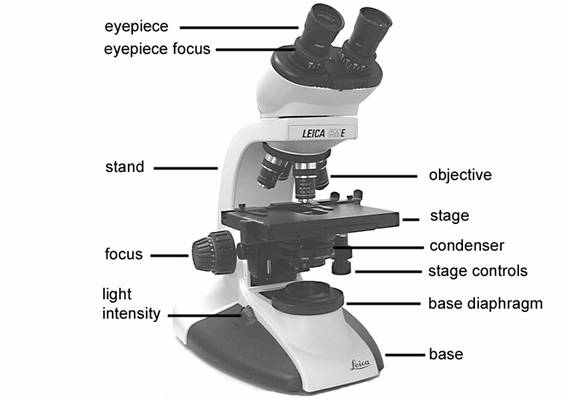
8 Best Images of Lens Diagram Worksheet - Microscope with Labeled Parts ...
Labelling the eye — Science Learning Hub Labelling the eye Add to collection The human eye contains structures that allow it to perceive light, movement and colour differences. In this activity, students use online or paper resources to identity and label the main parts of the human eye. By the end of this activity, students should be able to: identify the main parts of the human eye

eye diagram - PurposeGames
Diagram of the Eye - Lions Eye Institute To understand the eye and its functions, it's important to understand how the eye works, see below diagrams for both the external eye and the internal eye. The External Eye Instructions Click the parts of the eye to see a description for each. Hover the diagram to zoom. The Internal Eye Instructions

Uveal Coloboma – Portland Eye Care | Optometrist | Eyeglasses | Contact ...
FREE! - Label the Eye Worksheet – Teacher-Made Learning Resources In this resource, you’ll find a 2-page PDF that is easy to download, print out, and use immediately with your class. The first page is a labelling exercise with two diagrams of the human eye. One is a view from the outside, and the other is a more detailed cross-section. On the second page, you’ll find a set of answers showing the properly labelled human eyes, designed to help you check ...

What Does Your Eye Color Say About You? - PositiveMed | Eye color facts ...
Nervous System Worksheet Answers - WikiEducator Jan 14, 2008 · 8. The diagram below shows a section of a dog’s brain. Add the labels in the list below and, if you like, colour in the diagram as suggested. Cerebellum - blue; Spinal cord - green; Medulla oblongata - orange; Hypothalamus - purple; Pituitary gland - red; Cerebral hemispheres – yellow. 9. Match the descriptions below with the terms in the list.
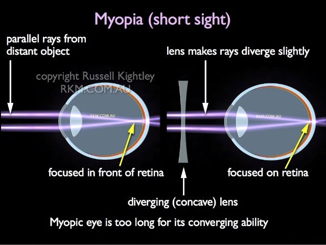
Vision defects and corrective lenses
Label Parts of the Human Eye Parts of the Eye Select the correct label for each part of the eye. The image is taken from above the left eye. Click on the Score button to see how you did. Incorrect answers will be marked in red.
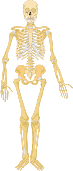
Skeleton Clip Art at Clker.com - vector clip art online, royalty free ...
The Eye - diagram to label | Teaching Resources File previews. pdf, 2.94 MB. Diagram of eye with key words to use in labelling it. Tes classic free licence.

Nitrogen Cycle Diagram to Label Inspirational Objectives Summarize the ...
eye labeling Diagram | Quizlet Middle layer of the eye (between the retina and the sclera) that contains the blood vessels that nourish the eye and cornea. iris. colored layer that dilates and constricts to allow in more or less light. ciliary body. structure on each side of the lens that connects the choroid and iris. Contains muscles that change the shape of the lens and ...

Football Pitch Soccers Field Measurements Clip Art at Clker.com ...
The Eyes (Human Anatomy): Diagram, Optic Nerve, Iris, Cornea ... - WebMD Articles On Eye Basics. Your eye is a slightly asymmetrical globe, about an inch in diameter. The front part (what you see in the mirror) includes: Iris: the colored part. Cornea: a clear dome ...
Post a Comment for "44 diagram of an eye with labels"